SUV measurement tools
Note: This feature requires a separate license and may not be available. Contact your system administrator to have this feature licensed and enabled.
SUV can be measured in a PET study in 2D mode or in Fusion mode. In Fusion mode, SUV measurements can be performed only on series that match the acquisition plane (typically axial). In SUITESTENSA, SUV can be measured only for BQML (becquerel / milliliter) units using any of the following measurement methods: body weight (bw), lean body mass (lbm), and body surface area (bsa). For more information on how these measurements are calculated, see SUV measurement methods.
Note that some essential data must be available in the DICOM header to perform SUV measurements (see Required DICOM attributes to calculate uptake values). If essential data is missing, the SUV measurement cannot be performed and the "SUV not available" indicator will display in the viewport with a tool tip that provides details about the missing information. If necessary, non-essential data such as sex, weight, height, dose, half life, radiopharmeceutical, and time of administration can be manually edited in the Advanced Visualization Parameters panel.
Important note on SUV calculations
SUV calculations in SUITESTENSA are based on industry-accepted standards and equations. They may differ from the methods of the customer. The customer is responsible for evaluating and approving the use of SUV measurements in their facility before the tool is approved for widespread use.
This section contains the following information:
- Overview of SUV measurement tools
- Change SUV parameters
- Change the SUV measurement method
- Measure SUV using an ROI tool
- Measure SUV using the Point Tool
- Save SUV measurements
- SUV measurement methods
Overview of SUV measurement tools
|
|
Circle ROI A single left-click starts the measurement and a second left-click ends the measurement. To edit, select the measurement to be active and then drag the measurement handles. The source of the calibration is shown in brackets at the bottom of the overlay. |
|
|
Ellipse ROI Left-click to start the measurement and left-click again to end the measurement. To edit a measurement, click the measurement to select it and then drag the measurement handles. The source of the calibration is shown in brackets at the bottom of the overlay. |
|
|
Rectangle ROI Left-click to start the measurement and left-click again to end the measurement. To edit a measurement, click the measurement to select it and then drag the measurement handles. The source of the calibration is shown in brackets at the bottom of the overlay. |
|
|
Freeform ROI Left-click to start the measurement and left-click on the first point selected or double-click to end the measurement. To edit a measurement, click the measurement to select it and then drag the measurement handles. The source of the calibration is shown in brackets at the bottom of the overlay. |
|
|
Point Tool Measures the exact value for a specific point. The unit of measure depends on the modality type. For example, in PET images it provides the SUV value, and in CT images it provides the Hounsfield units. Note that when using the Point Tool on reconstructed series, measurements can be made only on series that match the acquisition plane (typically axial). |
Change SUV parameters
If non-essential parameters such as patient weight or administration time are entered incorrectly at the time of acquisition, you can manually update these parameters in the Advanced Visualization Parameters panel and optionally save them as a Presentation State.
- On the Fusion tab, click
 to open the Advanced Visualization Parameters panel.
 - In the panel, click the SUV Parameters tab.
- Change any of the non-essential parameters such as sex, weight, height, dose, half life, radiopharmeceutical, and time of administration.
- Click Apply to apply your changes to the current view.
- Click Cancel to discard any un-applied changes.
- Click
 on the Home tab to reset all parameters back to the values you started with (if it is the original study, the values are set back to the DICOM values, and if it is a Presentation State, the values are set back to the original Presentation State values).
 - Click Reset (to the right of the associated parameter) to reset the parameter's value back to the original DICOM value (even in a Presentation State, this will revert to the original DICOM value).
- To save the parameters that you have changed, click Apply to apply the changes and then save the study as a Presentation State. Note that the study must also contain markup on the 2D PET (for instance an ROI measurement) to be able to save it as a Presentation State. For more information, see Save SUV measurements .
In the Advanced Visualization Parameters panel, a missing parameter displays a yellow warning marker and a modified parameter displays a checkmark beside it. Updated parameters are also shown in the SUV measurement overlay as shown in the image below. The units of measure, shown on the first line of the SUV overlay, indicate that they are based on "updated" parameters and the changed parameters are listed in the last line of the SUV overlay.
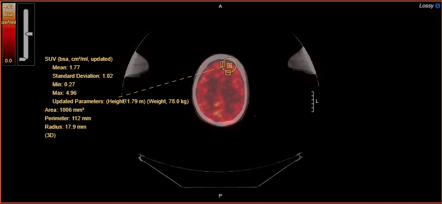
Figure 1 : Updated height and weight parameters in SUV measurement overlay
Change the SUV measurement method
The measurement method and the units are shown in the SUV measurement overlay. The available measurement methods and units are:
- gram per milliliter (g/ml) for body weight (bw) and lean body mass (lbm)
- centimeter-squared per milliliter (cm2/ml) for body surface area (bsa)
- On the Fusion tab, click and click the SUV Parameters tab.
- Select a measurement method.
- Click Apply.
Measure SUV using an ROI tool
When you measure SUV using an ROI tool, the SUV overlay shows the average and standard deviation as well as the minimum and maximum pixel values within the boundaries of that ROI. System administrators can configure what is shown in the overlay.
- On the Markup tab, click any of the ROI measurement tools or click the Circle ROI tool from the Fusion tab.
- Click and drag to outline the region of interest. (In the case of the polygon ROI, double-click to end the measurement.) Note that in Fusion mode, SUV measurements can be made only on series that match the acquisition plane (typically axial).
- The SUV values are displayed in the measurement overlay.
Measure SUV using the Point Tool
The Point Tool measures the exact SUV value for a specific point.
- On the Fusion tab, click

- Click the location that you want to measure. Note that in Fusion mode, SUV measurements can be made only on series that match the acquisition plane (typically axial).
Save SUV measurements
SUV measurements that are made on the original 2D axial PET images can be saved as a Presentation State. If any SUV parameters have been changed and those changes have been applied to the study, they will be saved as part of the Presentation State.
- On a 2D axial PET image, measure SUV using an ROI tool.
- To save a Presentation State, click
 on the study pill in the Series Tray.
 - In the confirmation dialog, select the markup that you want to save and click Yes.
SUV measurement methods
As mentioned earlier, SUITESTENSA supports the following measurement methods: body weight (bw), lean body mass (lbm), and body surface area (bsa). This section explains the calculation details for each of these methods.
Note on SUV measurements in Fusion images
Since fusion images are constructed from a volume, SUV computation is subject to interpolation algorithms.
Required DICOM attributes to calculate uptake values
The following DICOM attributes are required to calculate uptake values.
|
DICOM attribute |
Alternative Name Used in This Documentation | Tag |
|---|---|---|
| Corrected Image | (0028,0051) | |
| Decay Correction | (0054,1102) | |
|
Radiopharmaceutical Information Sequence
|
Radiopharmaceutical Injected Dose Radiopharmaceutical Administration Time |
(0054,0016) (0018,1074) (0018,1075) (0018,1072) (0018,1078) |
|
Series Date Series Time |
Scan Time |
(0008,0021) (0008,0031) |
|
Acquisition Date Acquisition Time |
Frame Acquisition Start Time |
(0008,0022) (0008,0032) |
|
Frame Reference Time |
|
(0054,1300) |
|
Actual Frame Duration |
Frame Duration |
(0018,1242) |
|
Patient’s Weight (in Kilogram) |
|
(0010,1030) |
|
Patient’s Size (in Meter) |
Patient’s Height |
(0010,1020) |
|
Patient’s Sex |
|
(0010,0040) |
|
Philips SUV Scale Factor |
Philips Body Weight SUV Scale Factor |
(7053,1000) |
|
Philips Activity Concentration Scale Factor |
|
(7053,1009) |
|
GE Scan Date and Time |
|
(0009,100d) |
PET exam background information
A PET exam starts by administering a certain dose of a radiopharmaceutical to a patient. A delay is necessary for patient’s metabolic processes to deliver the radiopharmaceutical to the target of the study. After this delay, the PET scan is performed. The result of the scan is a series of PET images. A scan is divided into multiple frames (or bed positions) and the data acquired in each frame is used to generate several slices which belong to that frame.
Decay correction calculation
The radiopharmaceutical decay is a continuous process throughout the exam. As a result, the pixel data obtained at different times during the exam may have different interpretation. A correction is necessary to unify this interpretation across the series; this correction is usually applied in two steps, one at the modality and one in the viewer application.
SUITESTENSA allows the calculation of SUV if the underlying series is corrected for decay with respect to the scan time at the modality. This condition is satisfied if (a) corrected image attribute contains the value DECY, and (b) decay correction attribute has the value START. Note that this correction does not account for the decay from the radiopharmaceutical administration time to the scan time. SUITESTENSA does correct for that decay as described below.
Denote by  the radiopharmaceutical injected dose at the administration time, and by
  the half-life of the radionuclide used in the radiopharmaceutical. The values of
  and
and  have the units Becquerel (Bq) and second, respectively. Also, denote by
  the time interval from the radiopharmaceutical administration time to the scan time. If the scan time is valid, the corrected dose at the scan time is given by:

Scan time calculation
The scan time is valid if it has a value in compliance with the DICOM specification, and if it precedes the acquisition time of any frame in the exam. When the series is post-processed after acquisition, the scan time is overwritten and hence no longer valid. The actual scan time can still be indirectly calculated if the following information is available:
- Acquisition date and time – The combination of acquisition date and time denote the frame acquisition start time
- Frame reference time – The time interval from the actual scan time to the average activity time. The average activity time for a frame is defined as the time instant within that frame where the instantaneous rate of radionuclide disintegration (or activity) is equal to the average activity over the frame.
- Frame duration – The time interval from the frame acquisition start time to the frame acquisition end time.
The procedure to indirectly calculate the scan time is as follows:
- Calculate the time interval from the frame acquisition start time to the average activity time as follows. Let the frame acquisition start time coincide with the time origin
 = 0. Denote by
  the number of radionuclides at time
  . We have
 - Subtract the value calculated in Step 1 from the frame reference time; the result is the length of the time interval from the actual scan time to the frame acquisition start time.
- Subtract the value calculated in Step 2 from the frame acquisition start time; the result is the actual scan time.
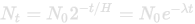
where  is known as the decay constant. The disintegration rate (or activity) at time
  is given by
  . From the definition of the average activity time, the interval of interest is the solution for t of the following equation:
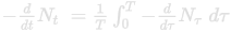
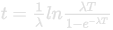
where  is the frame duration. The solution is given by:
SUV measurement calculations
The SUV is a normalized measure of radioactivity concentration and has the following generic form:

In the above formula,  is the normalization factor and
  is the activity concentration.
  is replaced with either the minimum or maximum pixel value within the boundaries of an ROI drawn over a PET or PET / CT fusion image, or the average value of all pixels within that ROI. The pixel values are directly read from the original full-resolution DICOM images before any transformation (such as Window Level) is applied.
We first consider a case where  has the unit BQML (Bq / Milliliter). In this case, the normalization factor can be defined in several ways.
Body weight SUV calculation
For body weight SUV, the normalization factor is the ratio of the patient’s weight  in gram to the corrected dose
  in Bq, i.e.,
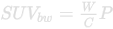
The SUV unit for body weight is gram / milliliter.
Lean body mass SUV calculations
For lean body mass SUV, the normalization factor is the ratio of the patient’s lean body mass in gram to the corrected dose  in Bq, i.e.,

The lean body mass in kilogram is calculated from the patient’s weight  in kilogram, height
  in centimeter and sex using James formula for females and Morgan formula for males:
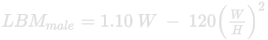
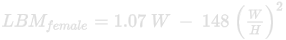
The SUV unit for lean body mass is gram / milliliter.
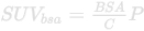
Body surface area SUV calulations
For body surface area SUV, the normalization factor is the ratio of the body surface area in centimeter-squared to the corrected dose  in Bq, i.e.,
The body surface area in centimeter-squared is calculated from the patient’s weight  in kilogram and height
  in centimeter using Du Bois formula:

The SUV unit for body surface area is centimeter-squared / milliliter.
Remarks
- Radiopharmaceutical administration date is implicitly inferred from radiopharmaceutical administration time, scan date and scan time. If the radiopharmaceutical administration time is before the scan time (e.g., radiopharmaceutical administration time = 14:27:00:000000 and scan time = 15:27:00:000000), it is assumed that the radiopharmaceutical administration date is the same as the scan date. If the radiopharmaceutical administration time is after scan time (e.g., radiopharmaceutical administration time = 23:29:00:000000 and scan time = 00:29:00:000000), it is assumed that the radiopharmaceutical administration date is one day before the scan date, i.e., the PET exam crosses midnight.
- Radiopharmaceutical start date time attribute is ignored. This DICOM attribute, if present, provides the explicit date on which the radiopharmaceutical is administered.
- We do not support GE private tag for scan date and time.
References
https://dicom.innolitics.com/ciods/pet-image/pet-image/00541300
http://qibawiki.rsna.org/images/8/86/SUV_vendorneutral_pseudocode_20180626_DAC.pdf